
Herpes Zoster Ophthalmicus Presenting with Suspected Orbital Myositis Following Recent Recombinant Zoster Vaccination
Abstract
Herpes zoster ophthalmicus (HZO) is a manifestation of varicella-zoster virus (VZV) reactivation involving the ophthalmic division of the trigeminal nerve, carrying significant risk of vision-threatening complications. Diplopia in HZO is often attributed to cranial nerve palsy, although orbital myositis remains a rare and underrecognized cause. We present a 79-year-old male who developed right-sided headache, binocular diplopia, and a V1 vesicular rash two weeks after receiving the recombinant zoster vaccine. Examination revealed restriction of extraocular movements without a localizing cranial nerve pattern, raising suspicion for orbital myositis. Neuroimaging was unremarkable, and VZV polymerase chain reaction confirmed the diagnosis of HZO. The patient received antiviral therapy and was discharged in stable condition after three days. This case highlights HZO presenting with suspected orbital myositis in temporal association with vaccination and underscores the need for vigilance for uncommon neuro-ophthalmic manifestations, as early recognition and treatment are essential to prevent vision-threatening complications. Given the patient's advanced age, this case also emphasizes the possibility that age-related immunosenescence may contribute to VZV reactivation and the development of HZO-related ocular complications. Potential therapeutic approaches targeting age-related immune dysfunction are also considered.
Author Contributions
Academic Editor: Ian James Martins, Principal Research Fellow, Edith Cowan University.
Checked for plagiarism: Yes
Review by: Single-blind
Copyright © 2026 Muhammad Awan, et al.
 This is an open-access article distributed under the terms of the Creative Commons Attribution License, which permits unrestricted use, distribution, and reproduction in any medium, provided the original author and source are credited.
Competing interests
The authors have declared that no competing interests exist.
Citation:
Introduction
Herpes zoster (HZ) results from the reactivation of latent varicella-zoster virus (VZV), which persists in the dorsal root and cranial nerve sensory ganglia following primary infection (i.e., chickenpox) 1. Reactivation risk increases with age or during episodes of illness or stress, largely due to declining cell-mediated immunity in a process known as immunosenescence. Herpes zoster clinically presents as a dermatomal vesicular rash that is often extremely painful and is typically preceded by a prodrome of localized burning or dysesthesia 2.
Herpes zoster ophthalmicus (HZO) involves the V1 (ophthalmic) branch of the trigeminal nerve and occurs in approximately 10-25% of all herpes zoster cases 3. Prompt antiviral treatment is crucial due to the potential involvement of the cornea, uvea, retina, and cranial nerves. Complications such as keratitis, uveitis, optic neuritis, or acute retinal necrosis are vision-threatening if not treated promptly 4.
Diplopia occurs in fewer than 30% of HZO cases and most often results from cranial nerve palsy involving the oculomotor (CN III), abducens (CN VI), or trochlear (CN IV) nerves, whether individually or in combination 5. Rarely, diplopia may result directly from orbital myositis, an inflammatory enlargement of the extraocular muscles. Orbital myositis can present with periorbital pain and edema, proptosis, diplopia, and ptosis 6. Recognition of these features is critical, as orbital myositis can precede the characteristic dermatomal rash, potentially delaying diagnosis and treatment 7.
The recombinant zoster vaccine (RZV, Shingrix), approved in 2017, is recommended for immunocompetent adults aged 50 and above. It is a non-live vaccine containing VZV glycoprotein E with an AS01B adjuvant, enhancing immune response without directly causing infection 8. While no causal relationship has been established between Shingrix and zoster onset shortly thereafter, there has been in increase in case reports noting a temporal association between the two 3.
We present a 79-year-old male who developed binocular diplopia and headache approximately two weeks after recombinant zoster vaccination and was subsequently diagnosed with HZO with suspected orbital myositis. This case highlights a rare clinical manifestation and a potential temporal association with recent vaccination, contributing to the growing body of literature on VZV reactivation following vaccination.
Case Presentation
A 79-year-old male with a history of hypertension, hyperlipidemia, and benign prostatic hyperplasia presented to the emergency department with a 10-day history of gradual-onset, low-grade right-sided headache and one day of new-onset diplopia. The headache was localized to the right forehead with occasional radiation to the right cheek. He described the pain as mild and persistent and denied any prior similar episodes. One day prior to presentation, he developed binocular diplopia that was most noticeable when looking at distant objects, which he described as vertically stacked images that resolved with monocular occlusion. He also noted the development of a rash over the right forehead. He denied any additional neurologic symptoms, including weakness, numbness, dysarthria, or gait instability. Of note, he had received his second dose of the recombinant zoster vaccine approximately two weeks prior to symptom onset.
His home medications included acetaminophen as needed, amlodipine 5 mg daily, losartan 50 mg twice daily, and rosuvastatin 5 mg nightly. He had no prior surgical history, denied tobacco or illicit drug use, and reported occasional alcohol use. He had no known drug allergies. On presentation, his blood pressure was 160/74 mmHg with otherwise normal vital signs. Physical examination revealed an evolving vesicular rash across the right forehead tender to palpation (Figure 1).
Figure 1.Unilateral vesicular rash across the right forehead tender to palpation.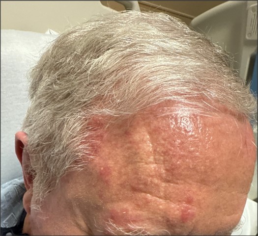
Neurologic examination was non-focal, and the patient was alert and oriented to person, place, and time. Cranial nerve evaluation revealed non-localizing binocular diplopia. Given the acute onset of diplopia, a stroke alert was activated on arrival. His National Institutes of Health Stroke Scale score was 0, and thrombolytic therapy was not administered. Initial laboratory evaluation, including complete blood count and basic metabolic panel, was largely within normal limits, with the exception of mild anemia and hyperchloremia (Table 1). Erythrocyte sedimentation rate (ESR) and C-reactive protein (CRP) were within normal limits, and hemoglobin A1c (HbA1c) was 5.8%.
Table 1. Complete blood count and basic metabolic panel results. “L” signifies values below the reference range; “H” indicates values above the reference range.| Test | Patient’s Value | Reference Values |
|---|---|---|
| White blood cell count (WBC) | 9.0 | 4.0-11.0 x 10³/μL |
| Hemoglobin (Hgb), male | 13.5 (L) | 14-18 g/dL |
| Hematocrit (Hct), male | 40.7 (L) | 42%-50% |
| Platelet (Plt) | 256 | 150-450 x 10³/μL |
| Sodium (Na) | 141 | 136-145 mEq/L |
| Potassium (K) | 3.9 | 3.5-5.0 mEq/L |
| Chloride (Cl) | 109 (H) | 98-106 mEq/L |
| Bicarbonate (CO₂) | 24 | 23-28 mEq/L |
| Anion Gap | 8 | 7-13 mEq/L |
| Blood Urea Nitrogen (BUN) | 15 | 8-20 mg/dL |
| Creatinine, male | 0.89 | 0.70-1.30 mg/dL |
| Glucose | 84 | 70-99 mg/dL |
| Osmolality | 290 | 275-295 mOsm/kg H₂O |
| Calcium | 8.8 | 8.6-10.2 mg/dL |
Reference ranges from the American Board of Internal Medicine 9.
Given concern for a neurologic etiology, the patient was admitted for further evaluation. Computed tomography (CT) of the head without contrast showed no evidence of mass, hemorrhage, midline shift, or extra-axial fluid collection, with clear paranasal sinuses and patent basilar cisterns. Magnetic resonance imaging (MRI) of the brain without contrast demonstrated no evidence of acute infarction or other intracranial pathology, with only minimal chronic microvascular changes noted. Carotid duplex ultrasound revealed no hemodynamically significant stenosis, and transthoracic echocardiogram showed no intra-atrial shunt or patent foramen ovale.
Ophthalmology was consulted due to concern for HZO involving the right V1 distribution. Ophthalmologic examination on day 2 demonstrated visual acuity of 20/40 in both eyes, with pupils equal, round, and reactive to light and intraocular pressures of 15 mmHg bilaterally. Visual fields were full. Extraocular movement testing showed reduced inferior and temporal motion of the right eye, while movements in the left eye were full. Slit lamp examination confirmed the vesicular rash across the right forehead and eyebrow, with otherwise normal anterior segment findings bilaterally, including clear corneas, deep and quiet anterior chambers, and trace nuclear sclerosis of the lenses. Fundus examination revealed normal optic discs with cup-to-disc ratios of 0.5, and normal macula, vessels, and peripheral retina bilaterally.
The diplopia was suspected to be secondary to orbital myositis given the absence of a localizing cranial nerve pattern. Empiric antiviral therapy with intravenous (IV) acyclovir (10 mg/kg every 8 hours) was initiated on day 2. Neurology was consulted and initially recommended MRI of the orbits with and without contrast; however, infectious disease consultation led to VZV polymerase chain reaction (PCR) testing, which returned positive. Orbital imaging was subsequently deferred. During hospitalization, the patient remained neurologically stable without symptom progression. His diplopia persisted but did not worsen. His hospital course was also notable for poorly controlled hypertension, for which amlodipine was increased to 10 mg daily, metoprolol tartrate 12.5 mg twice daily was initiated, and losartan 50 mg twice daily was continued.
The patient was discharged home in stable condition after a three-day hospitalization to complete a 14-day course of valacyclovir 1 g twice daily. He was instructed to follow up with his primary care physician within one to two weeks for blood pressure management and with his ophthalmologist for close monitoring of HZO and associated diplopia.
Discussion
HZ results from reactivation of latent VZV residing in sensory nerve ganglia, with risk factors including advanced age (particularly older than 60 years), immunosuppression, hypertension, and diabetes mellitus 10. HZ most frequently affects the thoracic dermatomes (approximately 50% of cases), followed by cranial nerve, cervical, and lumbar dermatomes (10-20%). Sacral dermatomes are least commonly affected (2-8%) 10.
The most common cranial nerve affected by VZV is the trigeminal nerve, specifically the ophthalmic division (V1). Involvement of the nasociliary branch produces vesicular lesions along the tip, side, or root of the nose - also known as the Hutchinson’s sign - which is associated with a higher risk of ocular complications 5. Up to 50% of patients with HZO develop ophthalmic complications, including diplopia, orbital myositis, cranial nerve palsies, keratitis, uveitis, and internuclear ophthalmoplegia. Diplopia may arise through two primary mechanisms, either through direct cranial nerve involvement or orbital inflammation affecting the extraocular muscles (i.e., orbital myositis) 5.
Diplopia secondary to cranial neuropathy can result from ischemic injury or direct viral-mediated inflammation. VZV may spread transaxonally along trigeminal ganglionic afferent fibers, reaching the vasa nervorum of adjacent cranial nerves and causing inflammation, ischemia, infarction, and demyelination 11. High-resolution MRI studies have demonstrated cavernous sinus enhancement extending to the cisternal segments of CN V, III, and VI in approximately 25% of patients with neuro-ophthalmic HZO, supporting a mechanism of direct inflammatory involvement rather than purely ischemic injury 12.
Cranial nerve palsies - most commonly involving CN III, IV, or VI - have been reported in up to 30% of patients with HZO and represent the more common etiology of diplopia in this setting. Diplopia due to cranial nerve palsies is typically painless 5. In contrast, orbital myositis, characterized by inflammation of one or more extraocular muscles, typically causes painful diplopia and restricted eye movements. Because both conditions may cause diplopia and limited extraocular movements, differentiating myogenic from neurogenic etiologies can be challenging without dedicated orbital imaging 12.
In our patient, the diplopia did not follow a pattern consistent with an isolated cranial nerve palsy. The restricted inferior and temporal motion of the right eye raised clinical suspicion for orbital myositis as the primary etiology. Clinical features suggestive of orbital myositis include mechanical restriction of extraocular movement rather than neurogenic weakness, involvement of multiple muscles simultaneously, pain with eye movement, and associated proptosis 4, 13. These findings support an inflammatory myopathic process rather than an isolated cranial neuropathy.
Orbital myositis is a rare and underdiagnosed complication of HZO, partly due to inconsistent use of orbital imaging in clinical practice and overlapping clinical features with cranial nerve palsies. A recent study using high-resolution MRI identified extraocular muscle inflammation indicative of myositis in 20 HZO patients confirmed by PCR, suggesting orbital myositis may be more prevalent in HZO than previously recognized 12. Recognition of this entity is critical, as it carries different prognostic implications from cranial neuropathy and may influence adjunctive management decisions.
Orbital myositis in HZO has a variable temporal relationship to the vesicular rash. It may present as a prodromal manifestation preceding cutaneous findings, concurrently with the rash, or as a delayed reaction appearing one to four weeks after skin involvement 14. When orbital myositis precedes the rash, the diagnostic window narrows if VZV is not considered early in the differential. Cases have been reported in which neuro-ophthalmic manifestations, including diplopia and periorbital pain, antedate cutaneous findings by several days, making a clinical diagnosis of HZO difficult to establish on clinical grounds alone 15, 16. VZV PCR testing of cutaneous vesicles, conjunctival swabs, or cerebrospinal fluid should therefore be pursued early when HZO is suspected, as confirmatory testing can expedite antiviral therapy and prevent further complications 4, 16.
Early initiation of antiviral therapy is the cornerstone of HZO management and should not be delayed when clinical suspicion is high, as prompt treatment is associated with reduced rates of ocular complications and postherpetic neuralgia. Both IV acyclovir and oral valacyclovir are effective; IV acyclovir is preferred in hospitalized patients with severe or sight-threatening disease at a dose of 10-15 mg/kg three times daily, with transition to oral valacyclovir upon stabilization 13. Our patient was started on IV acyclovir 10 mg/kg every 8 hours and discharged with a 14-day course of valacyclovir 1 g twice daily, consistent with favorable outcomes reported in HZO-associated orbital myositis 13. Patients typically experience gradual recovery over several months, though the timeline and extent of recovery vary.
The role of corticosteroids in HZO-associated orbital myositis remains incompletely defined, and antiviral therapy is generally considered the primary treatment. A systematic review and meta-analysis found that corticosteroid timing did not correlate with extraocular muscle recovery, whereas longer acyclovir duration was associated with significantly higher odds of recovery, suggesting antiviral duration may be the more impactful factor 17. Corticosteroid use in viral etiologies carries the theoretical risk of facilitating viral replication without adequate antiviral coverage 18. MRI of the orbits with and without contrast remains the preferred modality for confirming orbital myositis and should be strongly considered when the diagnosis is uncertain, intracranial extension is suspected, or the patient fails to respond to initial therapy 13. In our case, orbital imaging was deferred following a positive VZV PCR result and the clinical course remained stable, highlighting that MRI, while diagnostically valuable, may not be necessary in all cases when HZO with suspected orbital myositis is otherwise established.
This case underscores the importance of maintaining clinical suspicion for HZO even in the setting of recent zoster vaccination. Shingrix is a non-live recombinant vaccine containing VZV glycoprotein E with the AS01B adjuvant system and is highly effective in reducing herpes zoster incidence and its associated complications 19. However, recent studies have identified a brief post-vaccination window following the first dose during which there may be a transient increase in herpes zoster risk (up to 11-fold in one study) within the first 21 days in adults aged 65 years or older, with no such increase observed in younger adults and overall risk reduced after the second dose 20. A retrospective cohort study of 16,408 patients also found an approximately 60% increased risk of HZO recurrence within 56 days after receiving Shingrix in patients with a prior HZO history 3.
The mechanism underlying this transient risk is not fully understood, but the AS01B adjuvant may induce a robust immune response that temporarily disturbs the immunologic equilibrium maintaining VZV latency, creating a brief window during which reactivation is more likely in predisposed individuals. Importantly, this phenomenon does not imply vaccine causation of chronic disease; completing both doses results in a 73% reduction in shingles and approximately 78% reduction in HZO specifically 20. Recent vaccination should therefore not exclude the diagnosis when an elderly patient presents with a V1-distribution rash alongside periorbital or neuro-ophthalmic symptoms, as the immunosenescent state of older adults contributes to increased susceptibility to reactivation regardless of vaccination status 3.
A potential contributor to the immunosenescence underlying this presentation is the age-related decline of sirtuin 1 (SIRT1). SIRT1 is a highly conserved nicotinamide adenine dinucleotide (NAD+)-dependent deacetylase that serves as a crucial regulator of cellular metabolism and homeostasis. It also provides protection against chronic inflammation by controlling the acetylation of nuclear factor kappa B (NF-κB) 21. Notably, the expression of SIRT1 decreases with age across multiple tissues 22. Diminished SIRT1 activity may therefore contribute to the impaired cell-mediated immunity that predisposes older adults to VZV reactivation.
Within the eye, SIRT1 is expressed throughout ocular tissues and has been associate with protection against oxidative stress-related ocular disorders, including cataract, age-related macular degeneration, uveitis, and retinitis 23. The age-related decrease in SIRT1 activity may not only promote viral reactivation through immunosenescence but also increase susceptibility to vision-threatening complications following HZO. These observations raise interest in the potential pharmacologic modulation of SIRT1 in HZO-associated orbital myositis. Small molecule SIRT1 activators have demonstrated protective effects against oxidative stress and NLRP3 inflammasome activation in experimental retinal models 24. Conversely, SIRT1 inhibitors have been shown to enhance Type I and Type II interferon signaling, thereby augmenting the antiviral immune response 25.
Despite these findings, the effect of SIRT1 activation or inhibition in patients with HZO-associated orbital myositis remains unclear, and further investigation is required before therapeutic considerations can be established. Finally, circulating SIRT1 has been proposed as a serum marker of age-related decline 26. The early measurement of plasma SIRT1 levels in older adults may therefore help identify individuals with greater immunosenescence who are at increased risk for VZV reactivation and subsequent ocular complications.
Conclusion
This case illustrates herpes zoster ophthalmicus presenting with binocular diplopia and restricted extraocular movements suggestive of orbital myositis, arising in temporal association with recent recombinant zoster vaccination. Our case reinforces that orbital myositis is a rare, underrecognized cause of diplopia in HZO that may accompany or precede the dermatomal rash, complicating early diagnosis. Recent vaccination should not exclude HZO in older adults, given age-related immunosenescence and a transient post-vaccination reactivation window. Early clinical suspicion, timely VZV PCR confirmation, and prompt antiviral therapy remain essential to limiting vision-threatening complications and optimizing recovery. Finally, this study highlights the need for further research into immunosenescence and its modulation, including the potential role of SIRT1 as a biomarker of age-related immune dysfunction and a therapeutic target in HZO-associated ocular complications.
References
- 1.Son H, Kim S, Kwon J, Jung K H, Lee S et al. (2025) Varicella-zoster virus-specific cell-mediated immune response kinetics and latent viral load depending on aging. , J Med Virol 97(10), 10-1002.
- 2.Patil A, Goldust M, Wollina U. (2022) Herpes zoster: a review of clinical manifestations and management. Viruses. 14(2), 192-10.
- 3.Walia A, Sun Y, Acharya N R. (2024) Risk of herpes zoster ophthalmicus recurrence after recombinant zoster vaccination. , JAMA Ophthalmol 142(3), 249-56.
- 4.Minor M, Gurnani B, Payne E. (2026) Herpes zoster ophthalmicus. In: Stat Pearls [Internet]. Treasure Island (FL): Stat Pearls Publishing;.
- 5.Kedar S, Jayagopal L N, Berger J R. (2019) Neurological and ophthalmological manifestations of varicella zoster virus. , J Neuroophthalmol 39(2), 220-31.
- 6.Onder O, Bilgin R R, Koskderelioglu A, Gedizlioglu M. (2016) Orbital myositis: evaluating five new cases regarding clinical and radiological features. Noro Psikiyatri Ars. 53(2), 173-7.
- 7.Pereira A, Zhang A, Maralani P J, Sundaram A N. (2020) Acute orbital myositis preceding vesicular rash eruption in herpes zoster ophthalmicus. , Can 55-3.
- 9. (2024) American Board of Internal Medicine. ABIM laboratory test reference ranges [Internet]. , Philadelphia (PA): ABIM;
- 10.Steinmann M, Lampe D, Grosser J. (2024) Risk factors for herpes zoster infections: a systematic review and meta-analysis. Infection. 52(3), 1009-26.
- 11.Gilden D, Cohrs R J, Mahalingam R, Nagel M A. (2009) Varicella zoster virus vasculopathies: diverse clinical manifestations, laboratory features, pathogenesis, and treatment. Lancet Neurol. 8(8), 731-40.
- 12.Espinoza S, Labetoulle M, Rousseau A. (2026) High-resolution MRI for early diagnosis of orbital and neurovascular involvement in herpes zoster ophthalmicus. , Am 10.
- 13.Pace E, Accardo G, Lupia T, Brizzi M F, Corcione S et al. (2024) Orbital myositis after herpes zoster ophthalmicus: a case report and a narrative review of the literature. , Pathogens 13(10), 832-10.
- 14.Bak E, Kim N, Khwarg S I, Choung H K. (2018) Case series: herpes zoster ophthalmicus with acute orbital inflammation. Optom Vis Sci. 95(4), 405-10.
- 15.Kawasaki A, Borruat F X. (2003) An unusual presentation of herpes zoster ophthalmicus: orbital myositis preceding vesicular eruption. , Am 136(3), 574-5.
- 16.Rissardo J P, Patel P, Fornari Caprara AL. (2024) Herpes zoster ophthalmicus presenting with orbital myositis prior to the appearance of vesicular lesions: a case report and mini-review of the literature. Med Int (Lond). 4-6.
- 17.Shahriari M, Ashrafi S, Hosseini Imeni SM, Mohammad Soleymani S, Esmaily H. (2025) Treatment efficacy in herpes zoster ophthalmoplegia: a systematic review and meta-analysis of case reports and series. Rev Med Virol. 35-4.
- 18.Khadamy J, Khademi P, Baharypour S. (2023) Accelerated recovery: the role of oral steroid in the management of herpes zoster ophthalmicus-related ophthalmoplegia. Cureus. 15-12.
- 19.Anderson T C, Masters N B, Guo A. (2022) Use of recombinant zoster vaccine in immunocompromised adults aged ≥19 years: recommendations of the Advisory Committee on Immunization Practices - United States. 71(3), 80-4.
- 20.Shetty A N, Hennessy D, Kattan G S, Ojaimi S, Clothier H J et al. (2025) Transient increased risk of shingles post-Shingrix vaccination: self-controlled case-series analysis. Clin Infect Dis. 81-5.
- 21.Rahman S, Islam R. (2011) Mammalian Sirt1: insights on its biological functions. , Cell Commun Signal 9, 10-1186.
- 22.Gong H, Pang J, Han Y. (2014) Age-dependent tissue expression patterns of Sirt1 in senescence-accelerated mice. Mol Med Rep. 10(6), 3296-3302.
- 23.Mimura T, Kaji Y, Noma H, Funatsu H, Okamoto S. (2013) The role of SIRT1 in ocular aging. Exp Eye Res. 116, 17-26.
- 24.Li M, Xu Q, Fan Q. (2024) Small molecule SIRT1 activators counteract oxidative stress-induced inflammasome activation and nucleolar stress in retinal degeneration. , IntImmunopharmacol 142, 113167-10.